The progressive stage of multiple sclerosis (MS) has been suggested to be related to the central nervous system compartmentalization of the inflammation. Bates et Al. have been investigated the role of leptomeningeal inflammation in driving cortical pathology, focusing on the lymphotoxin (LTα) pathways that are known to play a key role in organogenesis and maintenance of lymphoid tissue. In detail, they show that:
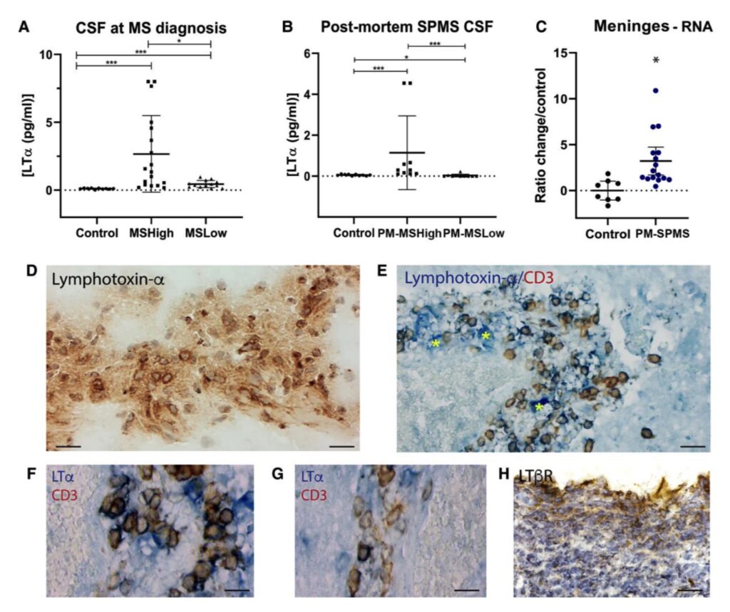
- LTα protein levels are higher in MS patients and in MS post-mortem brains respect to the controls especially in presence of multiple cortical lesions
- Recombinant hLTα injected into the subarachnoid space of the rat causes a transient infiltration of immune cells into the meninges and only in the rat immunized with MOG leads to subpial demyelination
- Injection of a LTα lentiviral vector into the cortical meningeal space (allowing the chronic effects of increased LTα expression to be studied) induces the formation of extensive meningeal aggregates of lymphoid-type immune cells, maintained over 3 months
- Injection of a LTα lentiviral vector into the cortical meningeal space also causes overexpression of lymphoid chemokines and extensive microglial-astroglial activation leading to subpial demyelination and marked neuronal loss resembling the typical cortical pathology of multiple sclerosis. Note that in these experiments the subpial demyelination is partially dependent on previous immunization with MOG, whereas the neuronal loss is present independently of immunization
In conclusion, this study demonstrates the relevant role of lymphotoxin pathways in determining the formation of meningeal lymphoid-like immune cell aggregates and multiple sclerosis-like cortical pathology, expanding our knowledge of the mechanisms underlying the progressive phase of multiple sclerosis.